The European network
for cell migration studies
PhD Student (project 4)
Biologist/Chemist
The position is part of the EU-funded Marie Skłodowska-Curie Innovative Training Network InCeM (Integrated Component Cycling in Epithelial Cell Motility). InCeM focuses on cell migration, which is essential for vital processes such as tissue formation, wound healing and tissue invasion during carcinogenesis. It aims to visualise morphological, biochemical and physical processes of cell motility to integrate these data into multi-scale models with the goal to deliberately tune motile behaviour in relation to disease. InCeM provides an international and highly interdisciplinary framework of collaborators from academia and industry with core expertise in medicine, biology, biochemistry, image analysis, modelling and engineering.
The topic of the subproject, which will be carried out at Uniklinik RWTH Aachen Institute of Molecular and Cellular Anatomy, is „Impact of keratin network regulation on migrating cells“. The epithelial cytoskeleton consists of keratin intermediate filaments together with actin filaments and microtubules, which form an integrated network whose dynamic organization is regulated by the extracellular matrix and adjacent cells through distinct adhesion sites. The project will employ printing techniques to systematically examine the effects of these adhesions and their related signalling mechanisms on cytoskeletal dynamics. Using novel image analysis tools comprehensive mathematical models will be developed in the partner laboratories of InCeM to predict migratory behaviour.
We are looking for a candidate with experience in molecular biology, microscopy techniques and functional assays. The candidate should have a strong background in cell biology and biophysics. We are looking for a motivated individual to pursue the highly interactive project toward a PhD degree.
PDF
Applications are invited for PhD student fellowships to start in spring/early summer 2015. The fellows should not have resided or carried out their main activity (work, studies, etc.) in the country of the host partner for more than 12 months in the 3 years immediately prior to the reference date. Salary details see PDF.
Application deadline: Applications should be submitted immediately but no later than the end of March to be considered and should be submitted using the application form.
For further enquiries concerning project details contact:
Prof. Dr. Rudolf E. Leube
InCeM Coordinator
Institute of Molecular and Cellular Anatomy
Uniklinik RWTH Aachen
Wendlingweg 2
52074 Aachen - Germany
This project has received funding from the European Union’s Horizon 2020 research and innovation programme under the Marie Sklodowska-Curie grant agreement No 642866.
NEWS
30th July 2015
InCeM
3.8 million euros grant success
for InCeM's MSCA-ITN-ETN
13th-31th July 2015
InCeM
In association with the Isaac Newton Institute:
Coupling Geometric PDEs with Physics for Cell Morphology, Motility and Pattern Formation
13th-24th July 2015
InCeM
Workshop Part 1 - Cambridge
InCeM Members at the Isaac Newton Institute in Cambridge during the 2. InCeM Workshop
GALLERY
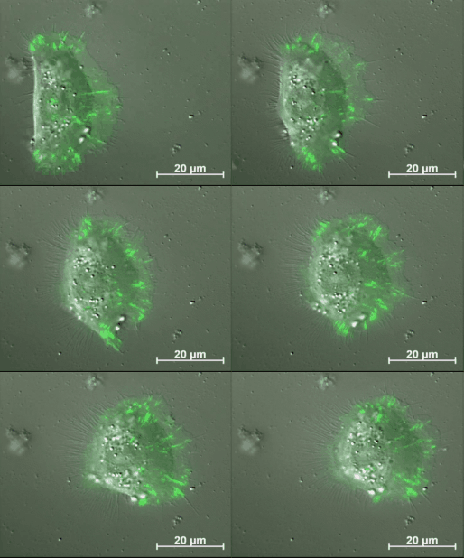
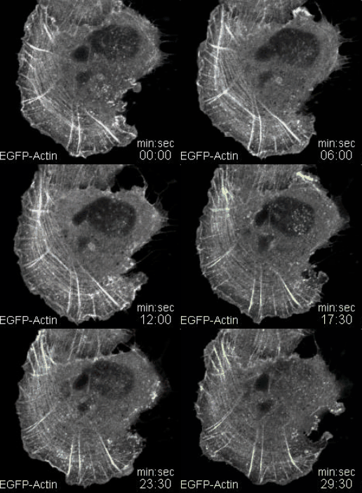
Migrating primary keratinocyte with labelled cell adhesions. Based on a fine tuned regulation adhesion structures are continuously formed, modified and finally disassembled.
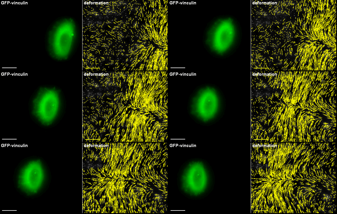
Primary keratinocytes apply traction forces upon migration. Using fluorescent fusion proteins (GFP-vinculin), adhesion structures can be visualized. Those sites are used by the cell for cell force transmission to the underlying substrate. In case those substrates are elastic, forces cause deformation fields that can be visualized using marker particles.
A typical example of the application of the evolving surface finite element method when solving partial differential equations of reaction-diffusion type on an evolving closed surface representing an evolving tumour.
A typical example of the application of the evolving surface finite element method when solving partial differential equations of reaction-diffusion type on an evolving open surface.
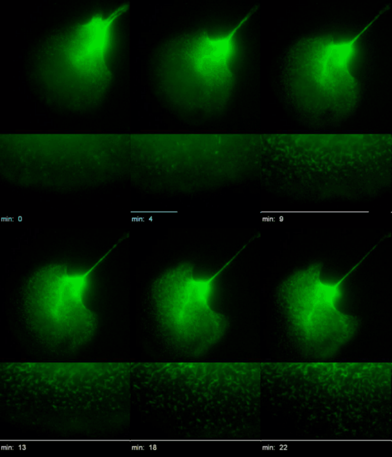
Time-lapse recordings of HK18-YFP fluorescence (left) of a migrating EK18-1 cell displaying multiple emerging KFPs in the proceeding lamellipodium. (Kölsch et al., 2010)
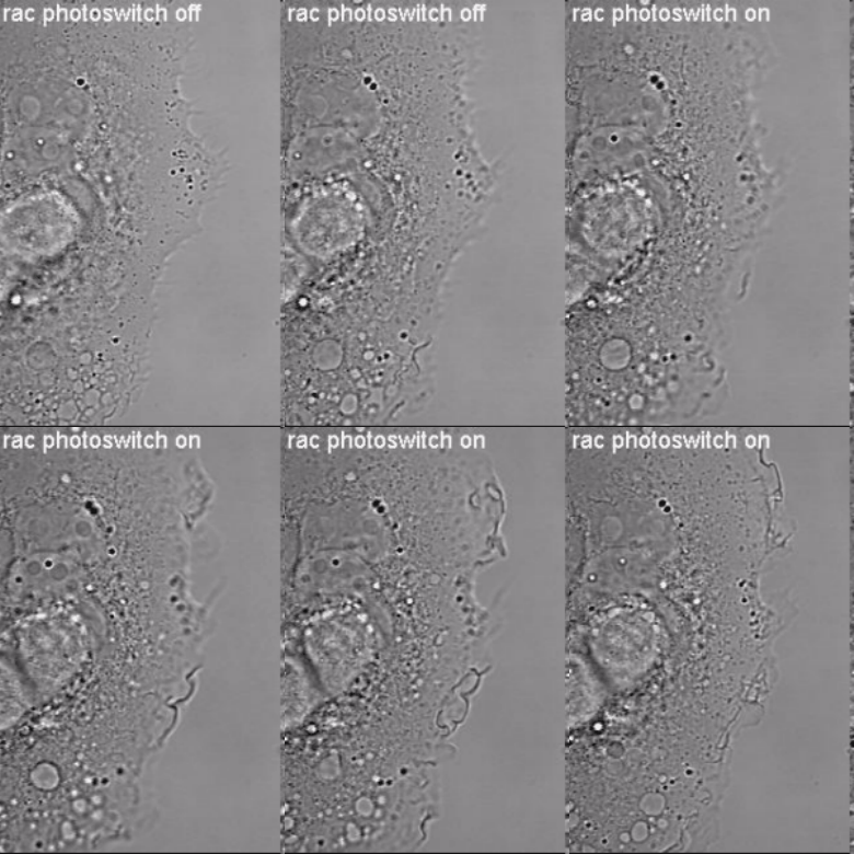
Induction of cell border dynamics through UV-light induced activation of pa-rac.
<
>
Update: 11.02.2016


