Workshop 2: Early start-up phase
3 weeks; target group: all ESRs and beneficiaries and partner organisations
This workshop will take place immediately after completing the recruitment phase:
Week 1 and 2:
Introduction to InCeM and setup of network structure (Isaac Newton Institute, Cambridge, UK)
- Introduction to the overall goals and rules of the training network and all of the partners (beneficiaries and partner organisations) with a brief description of the needs and concerns of InCeM. Presentation of the individual projects and training offers.
- Introduction to the field of cell migration research through introductory lectures on adhesion, migration, regulatory pathways, experimental techniques, image processing routines and different types of modelling approaches.
- Discussion and implementation of the rules for standardising experimentation (e.g., culture conditions, recording parameters), data management (e.g., storage formats, repositories, statistical methods, model outputs), modelling (calibration, validation, sensitivity and uncertainty analyses) and means of communication (wikis, homepages, online conferences).
- Establishment of working groups on (i) cytoskeletal organisation and dynamics during cell migration, (ii) regulatory mechanisms of cell migration, (iii) imaging and image processing, (iv) multidimensional data analysis and modelling and (v) tool development.
- Workshop in Cambridge
Week 3:
Introduction to cutting-edge techniques for examining and analysing cell migration
(FZJ Jülich/Uniklinik RWTH Aachen)
- Experimental techniques and devices in cell migration research.
- Image processing of dynamic structures
- Mathematical modelling of the cell.
Last update: 26.09.2018
incem@rwth-aachen.de
NEWS
January, 2019
InCeM
End of the InCeM Project
December 2018
InCeM
Last and final deliverable D6.4
“PhD theses” published!
November 2018
InCeM
A new publication of partner Forschungszentrum Jülich
GALLERY
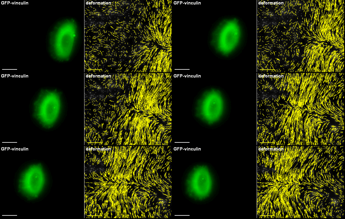
Migrating primary keratinocyte with labelled cell adhesions. Based on a fine tuned regulation adhesion structures are continuously formed, modified and finally disassembled.
Primary keratinocytes apply traction forces upon migration. Using fluorescent fusion proteins (GFP-vinculin), adhesion structures can be visualized. Those sites are used by the cell for cell force transmission to the underlying substrate. In case those substrates are elastic, forces cause deformation fields that can be visualized using marker particles.
A typical example of the application of the evolving surface finite element method when solving partial differential equations of reaction-diffusion type on an evolving closed surface representing an evolving tumour.
A typical example of the application of the evolving surface finite element method when solving partial differential equations of reaction-diffusion type on an evolving open surface.
Time-lapse recordings of HK18-YFP fluorescence (left) of a migrating EK18-1 cell displaying multiple emerging KFPs in the proceeding lamellipodium. (Kölsch et al., 2010)
Induction of cell border dynamics through UV-light induced activation of pa-rac.
<
>
This project has received funding from the European Union’s Horizon 2020 research and innovation programme under the Marie Sklodowska-Curie grant agreement No 642866.


