


6 - Life cycle analysis of actin, focal adhesions and force measurements
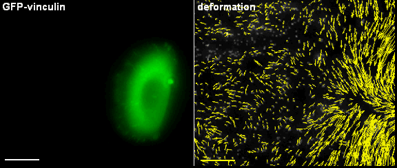
Primary keratinocytes apply traction forces upon migration. Using fluorescent fusion proteins (GFP-vinculin), adhesion structures can be visualized. Those sites are used by the cell for cell force transmission to the underlying substrate. In case those substrates are elastic, forces cause deformation fields that can be visualized using marker particles.
The aim of the project is to understand the mechanical function of specific actin filaments as one of the main structural cytoskeletal components of the cell. Using high-end microscopy techniques combined with laser cutting system we will analyze assembly, stabilization and disassembly of actin SFs, FAs and force generation with high spatiotemporal resolution during cell migration in primary keratinocytes (NHEK) and cell lines. Actomyosin force generation and transmission at FAs will be studied by life cell analyses on microstructured elastomeric substrates embedded with fluorescent beads. Results at the single cell level and averaged data will be incorporated into cell migration models and simulations. They will be applied to determine the interplay between force generation and assembly/disassembly of migratory structure components.
Last update: 28.05.2018
incem@rwth-aachen.de
Advanced cell migration assays (P1)
Chemotaxis and 2D/3D Migration (P2)
Analysis of keratin dynamics during migration (P3)
Impact of keratin network regulation on migrating cells (P4)
Correlation analyses of migration structure components and front-rear interplay (P5)
Life cycle analysis of actin, focal adhesions and force measurements (P6)
Monitoring of cancer cell migration in living animals (P7)
Principles of the filopodia structure, dynamics and mechanics (P8)
Mechanisms of downstream signalling from the Rho GTPase network to
cell morphogenesis and cell motility (P9)
Real-time tracking of keratinocyte migration and analysis of cell membrane shape changes (P10)
Image analysis of integrated cytoskeletal network dynamics (P11)
Coupling bulk-surface models for cell migration (P12)
Shaping membranes and actin fibres by forces (P13)
Integrating shape change models and imaging – inverse problem solving and model validation (P14)
Understanding spatio-temporal dynamics of the cytosol network during cell migration (P15)
This project has received funding from the European Union’s Horizon 2020 research and innovation programme under the Marie Sklodowska-Curie grant agreement No 642866.