Uniklinik RWTH Aachen (RWTH) - Germany
Supervisors: Rudolf Leube, Reinhard Windoffer, Alexander Bershadsky, Jacco van Rheenen, Marc Spehr
Student: Nadieh Kuijpers
3 - Analysis of keratin dynamics during migration

We are interested in the role of keratin intermediate filaments during cell motility and the interdependence between keratin and the other cytoskeletal compounds, such as actin and microtubules. Keratin cycling in migrating cells and sessile cell will be quantified using image analysis tools. These values can be used for mathematical modeling of the transition from non-migratory to migratory. Chemical and genetic manipulations will be used in combination with novel imaging techniques to reconstruct the interactions between the cytoskeletal components. Together, this will lead to a better understanding in the cooperation between these components, needed for the highly regulated process of cell migration.
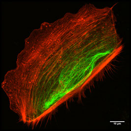
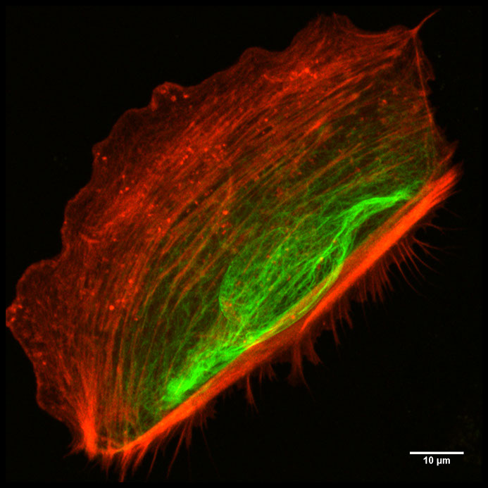
Maximum projection of actin (LifeAct-mRFP) and keratin intermediate filaments (K5-YFP) in a migrating nHEK cell.
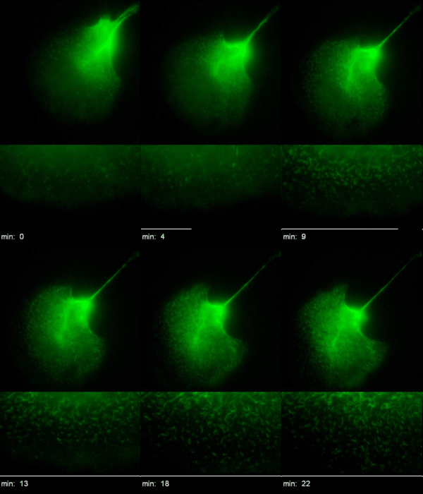
Time-lapse recordings of HK18-YFP fluorescence (left) of a migrating EK18-1 cell displaying multiple emerging KFPs in the proceeding lamellipodium. (Kölsch et al., 2010)
Last update: 28.05.2018
incem@rwth-aachen.de
Advanced cell migration assays (P1)
Chemotaxis and 2D/3D Migration (P2)
Analysis of keratin dynamics during migration (P3)
Impact of keratin network regulation on migrating cells (P4)
Correlation analyses of migration structure components and front-rear interplay (P5)
Life cycle analysis of actin, focal adhesions and force measurements (P6)
Monitoring of cancer cell migration in living animals (P7)
Principles of the filopodia structure, dynamics and mechanics (P8)
Mechanisms of downstream signalling from the Rho GTPase network to
cell morphogenesis and cell motility (P9)
Real-time tracking of keratinocyte migration and analysis of cell membrane shape changes (P10)
Image analysis of integrated cytoskeletal network dynamics (P11)
Coupling bulk-surface models for cell migration (P12)
Shaping membranes and actin fibres by forces (P13)
Integrating shape change models and imaging – inverse problem solving and model validation (P14)
Understanding spatio-temporal dynamics of the cytosol network during cell migration (P15)
This project has received funding from the European Union’s Horizon 2020 research and innovation programme under the Marie Sklodowska-Curie grant agreement No 642866.


