


Forschungszentrum Jülich GmbH (FZJ) - Germany
Student: Kritika Sahni
Supervisors: Rudolf Merkel, Bernd Hoffmann, Leif Dehmelt, Christian Thirion
5 - Correlation analyses of migration structure components and front-rear interplay
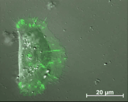
Migrating primary keratinocyte with labelled cell adhesions. Based on a fine tuned regulation adhesion structures are continuously formed, modified and finally disassembled.
Epithelial cell migration is being studied extensively over past few years. The process is described by a meshwork of interwoven course of actions pushing the leading edge forward and pulling on the rear end. The basal mechanism is been analyzed in detail and includes continuous assembly of various macromolecular protein complexes such as actin filaments, focal adhesions, etc. The molecular assembly takes place at the cell border, retrograde flow and migration of the cell itself leads to transfer of these molecules towards the center of the cell where they are finally disassembled. In a polarized cell, some molecules show very specific localized functions at the leading and at the rear end. How this molecular distribution takes place and what are the mechanisms behind the regulated distribution from the leading and the rear end still remains a big question. Considering these scientific questions the study involves liposomal vehicles to incorporate unspecific markers and specific proteins into a migrating cell. Molecules will be encapsulated into the fusion inducing liposomal carriers and delivered onto different cell sites. Following membrane fusion, molecules will be sorted to their natural locations and later their distribution behavior will be studied in 2D as well as in 3D.
Furthermore, the transport of different macromolecules within a migrating cell will be studied using photoswitchable fluorescent tags. A fusion protein construct will be formed where the molecule of interest will be coupled with a photoswitchable fluorescent protein on its preferred terminal site. Photoconversion on specific regions inside the cytoplasm/ membrane will enable tracking down the molecular movement in a migrating cell.
The ultimate aim of the study is to unravel the diffusion pattern or the active transport of molecules in cytoplasm or in the membrane both in 2D and in 3D.
Last update: 28.05.2018
incem@rwth-aachen.de
Advanced cell migration assays (P1)
Chemotaxis and 2D/3D Migration (P2)
Analysis of keratin dynamics during migration (P3)
Impact of keratin network regulation on migrating cells (P4)
Correlation analyses of migration structure components and front-rear interplay (P5)
Life cycle analysis of actin, focal adhesions and force measurements (P6)
Monitoring of cancer cell migration in living animals (P7)
Principles of the filopodia structure, dynamics and mechanics (P8)
Mechanisms of downstream signalling from the Rho GTPase network to
cell morphogenesis and cell motility (P9)
Real-time tracking of keratinocyte migration and analysis of cell membrane shape changes (P10)
Image analysis of integrated cytoskeletal network dynamics (P11)
Coupling bulk-surface models for cell migration (P12)
Shaping membranes and actin fibres by forces (P13)
Integrating shape change models and imaging – inverse problem solving and model validation (P14)
Understanding spatio-temporal dynamics of the cytosol network during cell migration (P15)
This project has received funding from the European Union’s Horizon 2020 research and innovation programme under the Marie Sklodowska-Curie grant agreement No 642866.